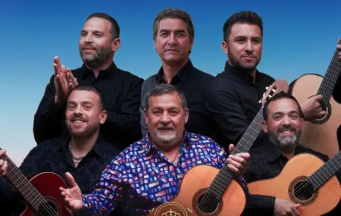
Gipsy Kings: "Music Is a Natural Way of Life"
Section: Arts
Recent advancements in organoid research have led to the successful development of organoids that contain blood vessels, offering significant potential for both scientific study and therapeutic applications. This innovative work, conducted by a team from Stanford Medicine, marks a pivotal step in overcoming previous limitations associated with organoid size and viability.
Organoids, which are miniature clusters of cells that replicate the function of specific organs, have been utilized in various fields of research. For example, brain organoids have been instrumental in exploring neurodevelopmental disorders, while intestinal organoids have been used to model conditions such as celiac disease. However, a major limitation has been their size; organoids are typically restricted to the dimensions of a sesame seed due to the absence of an internal blood vessel system that delivers essential nutrients and oxygen to all cells.
Oscar Abilez, a senior scientist at Stanford, highlighted the challenges faced when organoids exceed a certain size, leading to cell death in the center of the structure due to insufficient nutrient absorption. The recent study published in Science demonstrates a breakthrough by integrating tiny blood vessels within heart and liver organoids, potentially allowing them to grow larger and more mature.
This vascularization enhances the practicality of organoids as biological models, facilitating their use in regenerative medicine. According to Joseph Wu, the senior author of the study, the inclusion of a vascular system could enable the transplantation of vascularized cardiac organoids derived from a patient's own stem cells to repair damaged heart tissue.
In their research, the team explored a variety of chemical conditions to optimize the growth of cardiac organoids capable of producing the necessary cell types for forming blood vessels. Through experimentation with 34 distinct recipes, they identified a successful method that yielded organoids with a robust network of endothelial cells, smooth muscle cells, and cardiomyocytes.
The resulting organoids exhibited a structure where blood vessels were clearly defined and resembled the capillary networks found in a developing heart. Remarkably, these organoids contained a diverse array of cell types, closely mirroring the cellular composition of a six-week-old embryonic heart.
This finding offers valuable insights into the early stages of human development, a period that presents ethical challenges for direct study in living subjects. The researchers conducted preliminary tests on the organoids using fentanyl, revealing that exposure to this potent opioid resulted in an increase in blood vessel formation, indicating potential implications for neonatal health.
The methodology established for vascularizing cardiac organoids can also be adapted for other organ types, such as the liver, further expanding the scope of research possibilities. Future investigations will focus on allowing the vascularized organoids to mature further and optimizing the growth conditions to incorporate additional cell types, including immune and blood cells, to better replicate the characteristics of adult organs.
This pioneering work not only enhances the understanding of organ development and function but also paves the way for innovative approaches to treat various diseases and injuries through regenerative therapies.
Section: Arts

Section: Fashion
Section: Travel

Section: Health Insurance
Section: News

Section: Politics

Section: Business
Section: Health

Section: Arts
Section: Business
Both private Health Insurance in Germany and public insurance, is often complicated to navigate, not to mention expensive. As an expat, you are required to navigate this landscape within weeks of arriving, so check our FAQ on PKV. For our guide on resources and access to agents who can give you a competitive quote, try our PKV Cost comparison tool.
Germany is famous for its medical expertise and extensive number of hospitals and clinics. See this comprehensive directory of hospitals and clinics across the country, complete with links to their websites, addresses, contact info, and specializations/services.
Didn't manage to get a ticket for Linkin Park? Or still not enough after the concert? Join us at CRASH on June 12th for our "IN THE END" Linkin Park Special + CORE NIGHT.All night long, we'll be playing Linkin Park's music, along with Nu Metal, Metalcore, and Alternative Rock from bands such as Limp...


No comments yet. Be the first to comment!