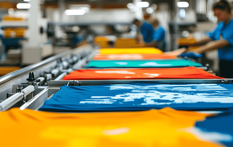
Custom T-Shirts: Last-Minute Gifts They'll Actually Love
Section: Arts
Recent developments in 3D bioprinting technology have ushered in new possibilities for personalized treatment of gastric cancer. Researchers from Pohang University of Science and Technology (POSTECH) have successfully created a gastric cancer model that utilizes patient-derived tissue fragments, preserving the essential characteristics of actual tumors. This groundbreaking model aims to enhance the speed and accuracy with which individual patient drug responses can be evaluated and predicted.
The challenge of tumor heterogeneity complicates cancer treatment, as patients often respond differently to the same therapeutic agents. Consequently, predicting the effectiveness of anticancer therapies is crucial in minimizing adverse side effects and improving overall treatment outcomes. Traditional methods, including gene panel testing and patient-derived xenograft (PDX) models, have limitations in their applicability and efficiency. They often require extensive time and resources to establish, which can hinder timely patient care.
In this innovative study, the research team developed an in vitro gastric cancer model by incorporating 3D bioprinting technology and a bioink made from tissue-specific materials that include patient-derived fragments. A significant aspect of this model is the encapsulation of cancer tissues within a decellularized extracellular matrix (dECM) hydrogel sourced from stomach tissues. This setup facilitates interactions between the cells and the matrix, effectively simulating the in vivo tumor microenvironment.
The newly developed model replicates the unique characteristics of gastric tissues from individual patients by mimicking critical interactions between cancer cells, their surrounding stroma, and the extracellular matrix. This leads to a high specificity in predicting responses to anticancer drugs, as well as prognostic outcomes. Moreover, the gene profiles associated with cancer progression and drug response closely align with those found in actual patient tissues, outperforming conventional PDX models.
Another significant advantage of this 3D bioprinting approach is its rapid fabrication process, allowing for drug evaluations to be conducted within two weeks of extracting tumor tissue from patients. This time-efficient model is expected to play a vital role in the advancement of personalized cancer therapies.
Professor Charles Lee, who leads the research, highlighted the potential of this model to enhance the precision of drug response predictions, thereby reducing the likelihood of administering ineffective treatments to patients who may not benefit from them. Meanwhile, Professor Jinah Jang from POSTECH emphasized the importance of this research as a critical preclinical platform, not only for developing tailored treatments but also for validating new anticancer drugs and potential combination therapies.
Section: Arts
Section: Arts

Section: Business

Section: Business
Section: Arts

Section: Health

Section: Arts
Section: News
Section: News
Section: Arts
Health Insurance in Germany is compulsory and sometimes complicated, not to mention expensive. As an expat, you are required to navigate this landscape within weeks of arriving, so check our FAQ on PKV. For our guide on resources and access to agents who can give you a competitive quote, try our PKV Cost comparison tool.
Germany is famous for its medical expertise and extensive number of hospitals and clinics. See this comprehensive directory of hospitals and clinics across the country, complete with links to their websites, addresses, contact info, and specializations/services.
One of the most beautiful squares transforms into a summer stage every year for two days. The Gärtnerplatz Open-Air features a free music and cultural program across three stages, as well as street food from local vendors. On Saturday, the main stage at Gärtnerplatz offers something for everyone,...


No comments yet. Be the first to comment!